Scientific Image Gallery
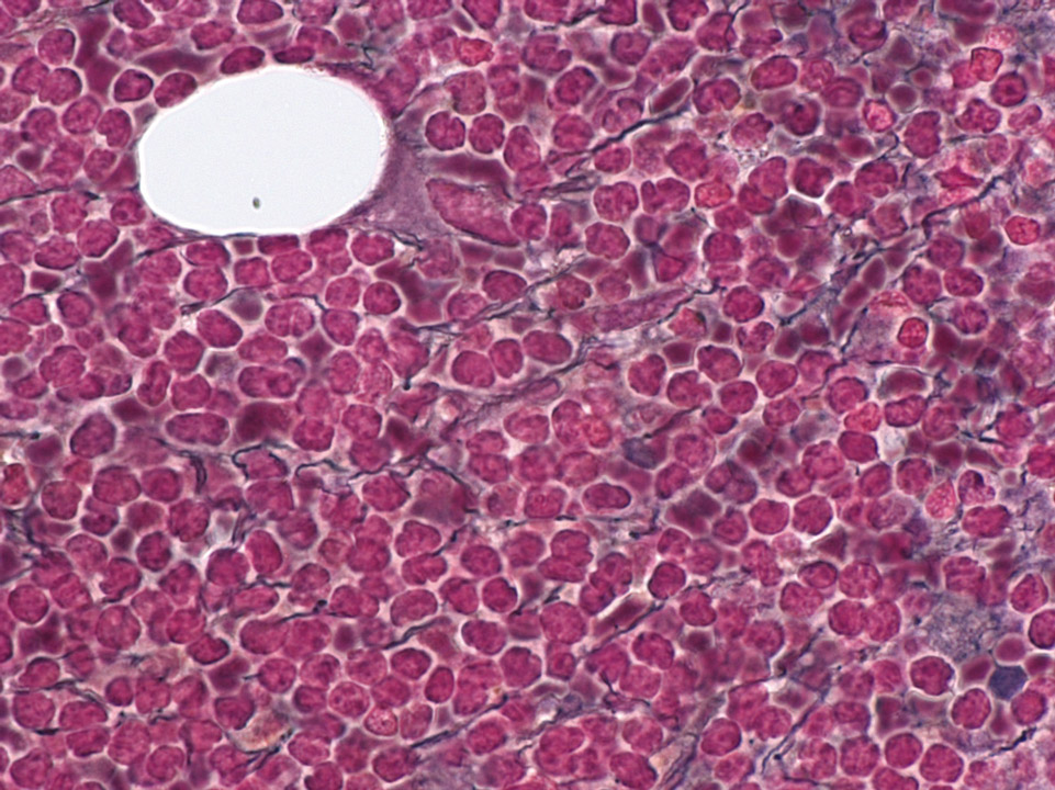
Bone marrow histology (Gomori stain) of a patient showing an infiltration of mantle cell lymphoma cells and an increase in fibres (black lines).
<p>Bone marrow histology (Gomori stain) of a patient showing an infiltration of mantle cell lymphoma cells and an increase in fibres (black lines).</p>
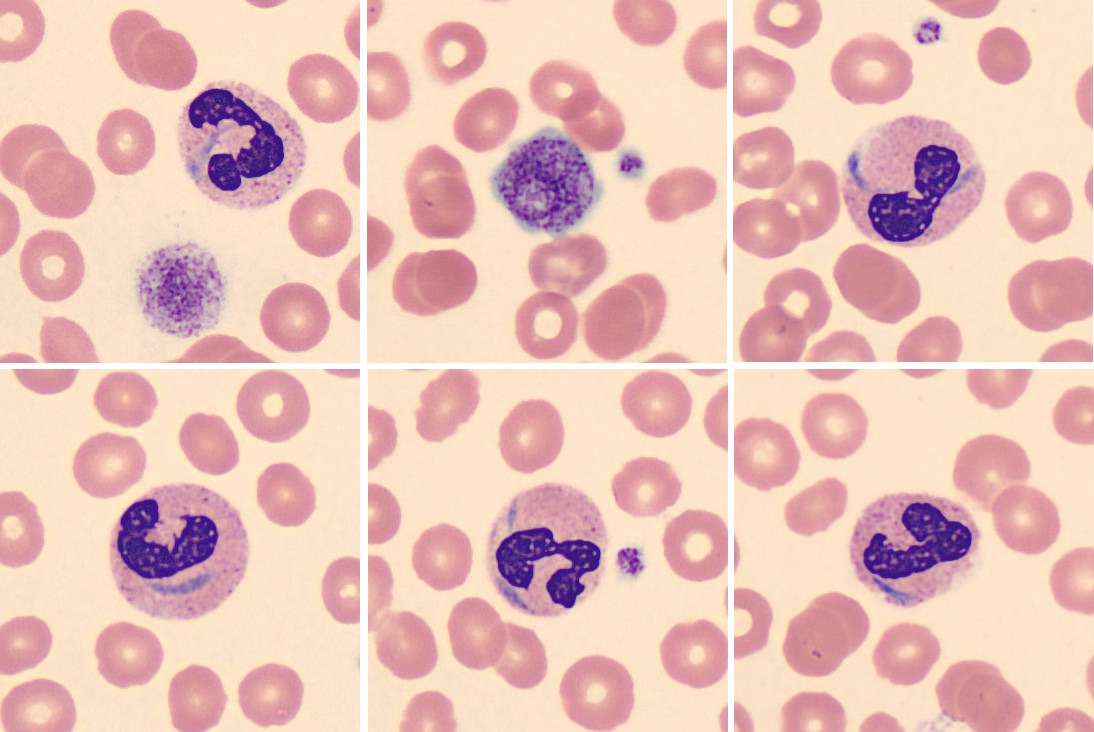
May-Hegglin anomaly belongs to a family of macrothrombocytopenias characterised by mutations in the MYH9 gene. It is a rare autosomal dominant disorder characterised by various degrees of thrombocytopenia that may be associated with purpura and bleeding.
<p>May-Hegglin anomaly belongs to a family of macrothrombocytopenias characterised by mutations in the MYH9 gene. It is a rare autosomal dominant disorder characterised by various degrees of thrombocytopenia that may be associated with purpura and bleeding. </p>
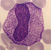
Cell description:
Size: 10-12 µm
Nucleus: kidney or U-shaped with clumped chromatin
Cytoplasm: acidophilic
neutrophil: fine reddish granulation
Cell division is not possible anymore and protein synthesis has stopped.
<p>Cell description: </p> <p>Size: 10-12 µm </p> <p>Nucleus: kidney or U-shaped with clumped chromatin </p> <p>Cytoplasm: acidophilic </p> <p>neutrophil: fine reddish granulation </p> <p>Cell division is not possible anymore and protein synthesis has stopped.</p>
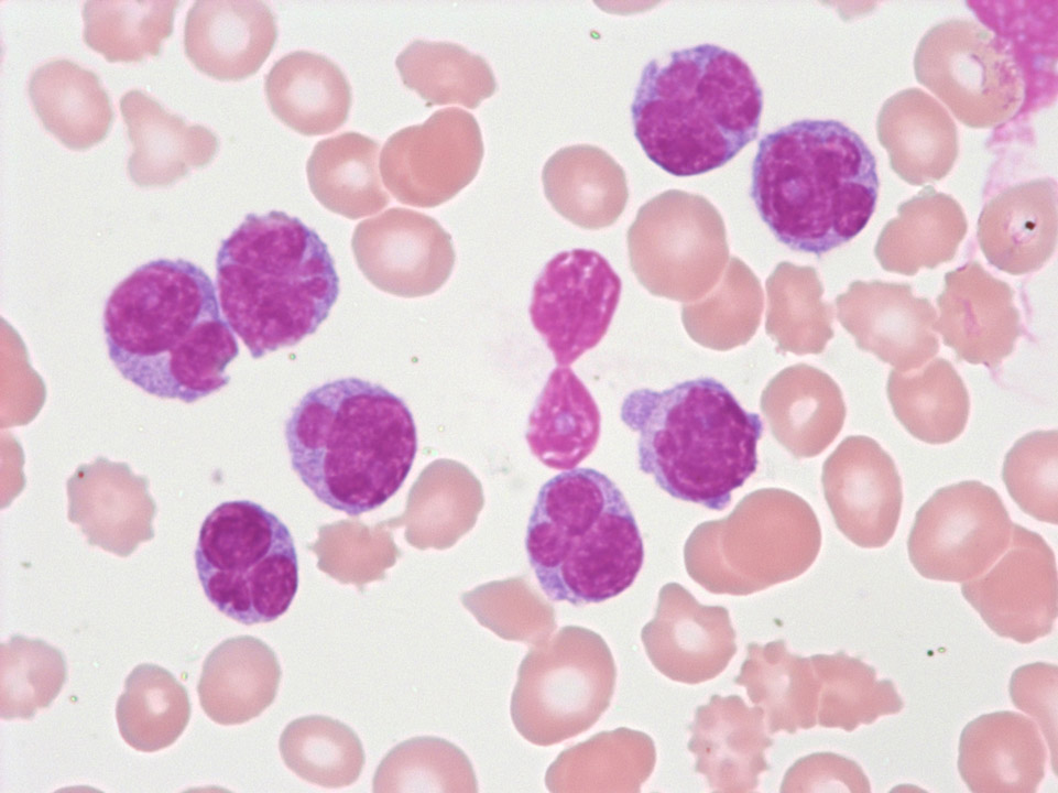
Blood film prepared after storage of the EDTA blood for more than one day. A safe morphological differentiation is no longer possible.
<p>Blood film prepared after storage of the EDTA blood for more than one day. A safe morphological differentiation is no longer possible.</p>
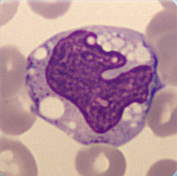
Cell description:
Size: 20 µm
Nucleus: kidney- to band-shaped
Cytoplasm: grey and clear with fine azurophilic granules
They are shortly located in the peripheral blood and then move into the tissue where they differentiate into macrophages. Function: Phagocytosis either of harmful pathogens or dead, dying or damaged cells from the blood.
<p>Cell description: </p> <p>Size: 20 µm </p> <p>Nucleus: kidney- to band-shaped </p> <p>Cytoplasm: grey and clear with fine azurophilic granules </p> <p>They are shortly located in the peripheral blood and then move into the tissue where they differentiate into macrophages. Function: Phagocytosis either of harmful pathogens or dead, dying or damaged cells from the blood. </p>
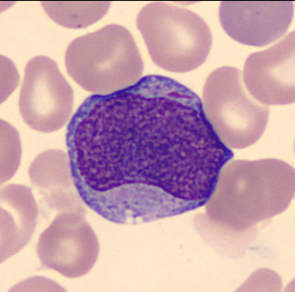
The first microscopically identifiable cell of granulocytic cell line.
Cell description:
Size: 12-20 µm
Nucleus: large, round or slightly oval with diffuse chromatin pattern and often 1-5 nucleoli
Cytoplasm: pale blue and usually agranular, sometimes Auer rods visible
<p>The first microscopically identifiable cell of granulocytic cell line. </p> <p>Cell description: </p> <p>Size: 12-20 µm </p> <p>Nucleus: large, round or slightly oval with diffuse chromatin pattern and often 1-5 nucleoli </p> <p>Cytoplasm: pale blue and usually agranular, sometimes Auer rods visible</p>
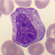
In this maturation stage the separation into the 3 different subpopulations of granulocytes occurs by development of specific granulation for each (secondary granulation).
Cell description:
Size: 10-18 µm
Nucleus: oval or slightly indented with variable degree of chromatin clumping, nucleoli usually not apparent
Cytoplasm: acidophilic neutrophil: primary azurophilic and secondary neutrophilic granules
<p>In this maturation stage the separation into the 3 different subpopulations of granulocytes occurs by development of specific granulation for each (secondary granulation). </p> <p>Cell description: </p> <p>Size: 10-18 µm </p> <p>Nucleus: oval or slightly indented with variable degree of chromatin clumping, nucleoli usually not apparent </p> <p>Cytoplasm: acidophilic neutrophil: primary azurophilic and secondary neutrophilic granules</p>
