Scientific Image Gallery

Auto-immune haemolytic anaemia (AIHA) in a case of chronic lymphocytic leukaemia (B-CLL): Reticulocytes are increased, spherocytes are present, Coombs' test is positive.
<p>Auto-immune haemolytic anaemia (AIHA) in a case of chronic lymphocytic leukaemia (B-CLL): Reticulocytes are increased, spherocytes are present, Coombs' test is positive.</p>

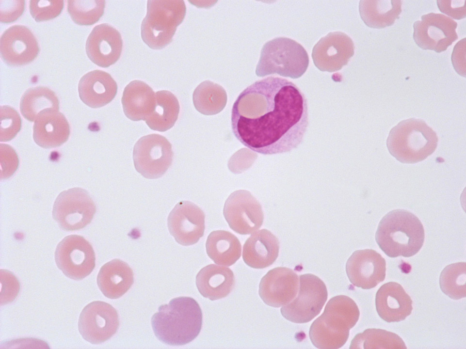
Phagocytosis of a red blood cell by a monocyte in a case of auto-immune haemolytic anaemia (AIHA). The many spherocytes and polychromasia are clearly visible.
<p>Phagocytosis of a red blood cell by a monocyte in a case of auto-immune haemolytic anaemia (AIHA). The many spherocytes and polychromasia are clearly visible.</p>

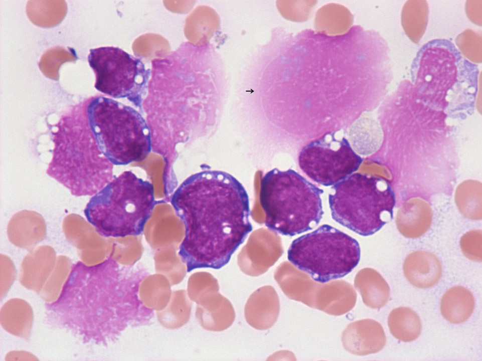
B-ALL/Burkitt lymphoma: The blasts typically show a deep blue cytoplasm and several vacuoles. Tumour cells tend to be very fragile, resulting in 'smudge' cells (->) during blood film preparation.
<p>B-ALL/Burkitt lymphoma: The blasts typically show a deep blue cytoplasm and several vacuoles. Tumour cells tend to be very fragile, resulting in 'smudge' cells (->) during blood film preparation.</p>
![[Sysmex WCA (english)] Basophil [Sysmex WCA (english)] Basophil](/fileadmin/media/f100/images/CellImages/Basophil.png)
Size: 10-14 µm
Nucleus: lobulated but often obscured
Cytoplasm: acidophil with purple-black granulation
Function: basophils can release histamine and heparin to respond to a suspected infection
<p>Size: 10-14 µm </p> <p>Nucleus: lobulated but often obscured </p> <p>Cytoplasm: acidophil with purple-black granulation </p> <p>Function: basophils can release histamine and heparin to respond to a suspected infection</p>

In a patient with CML the BCR-ABL fusion gene could be detected by fluorescence-in-situ-hybridisation (FISH, interphase preparation).
<p>In a patient with CML the BCR-ABL fusion gene could be detected by fluorescence-in-situ-hybridisation (FISH, interphase preparation).</p>


