Scientific Image Gallery
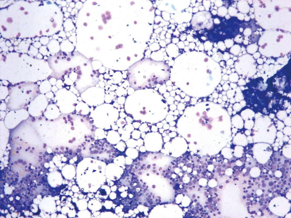
Bone marrow cytology (May-Grünwald-Giemsa stain) of a 37-year old patient with aplastic anaemia, showing a reduced cell content: In the upper half of the picture only fat cells are present, in the lower third haematopoietic cells are still present.
<p>Bone marrow cytology (May-Grünwald-Giemsa stain) of a 37-year old patient with aplastic anaemia, showing a reduced cell content: In the upper half of the picture only fat cells are present, in the lower third haematopoietic cells are still present.</p>
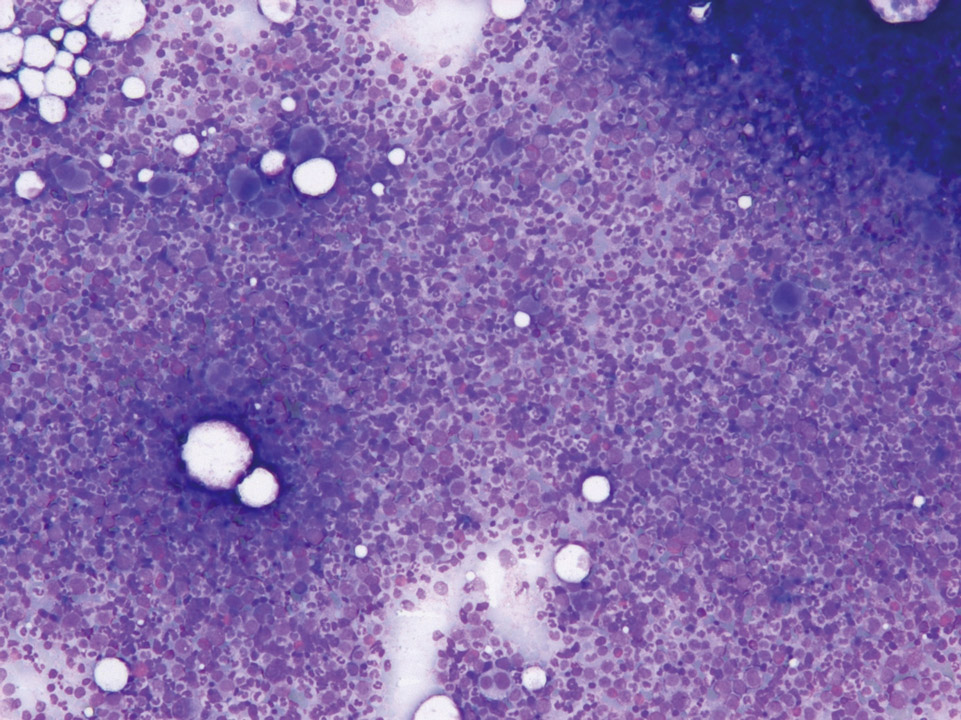
Bone marrow cytology (May-Grünwald-Giemsa stain) of a patient with CML demonstrating an extremely elevated number of cells: Granulopoiesis is enhanced, megakaryocyte count is increased. Cytogenetic detection of the Philadelphia chromosome (Ph1) confirmed the diagnosis of CML.
<p>Bone marrow cytology (May-Grünwald-Giemsa stain) of a patient with CML demonstrating an extremely elevated number of cells: Granulopoiesis is enhanced, megakaryocyte count is increased. Cytogenetic detection of the Philadelphia chromosome (Ph1) confirmed the diagnosis of CML.</p>
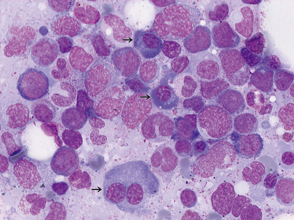
This bone marrow cytology (May-Grünwald-Giemsa stain) of a 75-year old patient with chronic myelomonocytic leukaemia (CMML) shows in addition a multiple myeloma with plenty of plasma cells (->).
<p>This bone marrow cytology (May-Grünwald-Giemsa stain) of a 75-year old patient with chronic myelomonocytic leukaemia (CMML) shows in addition a multiple myeloma with plenty of plasma cells (->).</p>
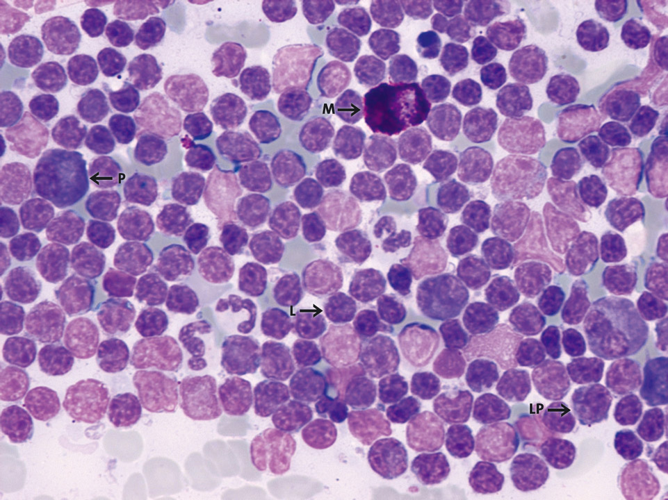
Bone marrow cytology (May-Grünwald-Giemsa stain) of a patient with immunocytoma showing a dense infiltration of small lymphocytes (L), lymphoplasmacytic cells (LP), a few plasma cells (P) and mast cells (M). The cells produce monoclonal IgM, characterising it as a Waldenström's disease.
<p>Bone marrow cytology (May-Grünwald-Giemsa stain) of a patient with immunocytoma showing a dense infiltration of small lymphocytes (L), lymphoplasmacytic cells (LP), a few plasma cells (P) and mast cells (M). The cells produce monoclonal IgM, characterising it as a Waldenström's disease. </p>
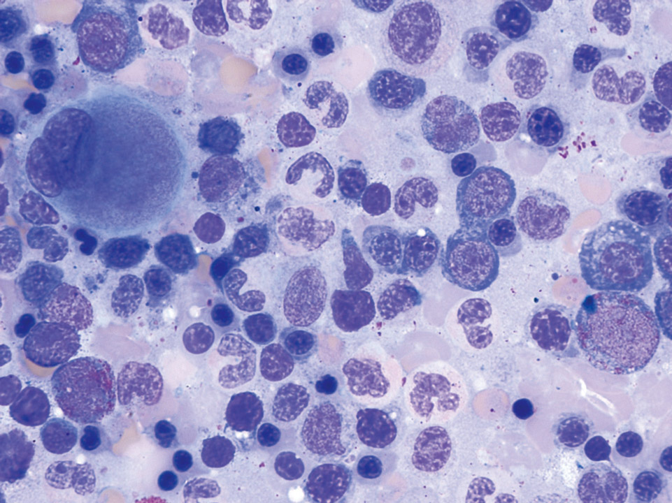
Bone marrow cytology (May-Grünwald-Giemsa stain) of an 82-year old patient, in which, considering the age of this patient, a substantial increase of cells and dysplastic changes in all cell lineages are evident. The percentage of blasts is below 5%. This is a refractory cytopenia with multilineage dysplasia (RCMD).
<p>Bone marrow cytology (May-Grünwald-Giemsa stain) of an 82-year old patient, in which, considering the age of this patient, a substantial increase of cells and dysplastic changes in all cell lineages are evident. The percentage of blasts is below 5%. This is a refractory cytopenia with multilineage dysplasia (RCMD).</p>
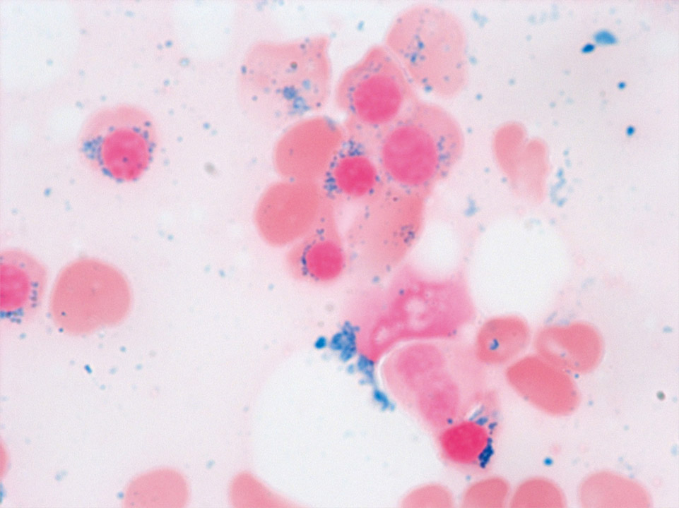
This bone marrow cytology of a patient with MDS shows predominantly ringed sideroblasts in the Prussian Blue stain (iron stain). They are precursors of red blood cells, in which, due to a defect in iron utilisation, the iron is deposited in a ring-shaped distribution around the nucleus. Exact diagnosis: refractory cytopenia with multilineage dysplasia and ringed sideroblasts (RCMD-RS).
<p>This bone marrow cytology of a patient with MDS shows predominantly ringed sideroblasts in the Prussian Blue stain (iron stain). They are precursors of red blood cells, in which, due to a defect in iron utilisation, the iron is deposited in a ring-shaped distribution around the nucleus. Exact diagnosis: refractory cytopenia with multilineage dysplasia and ringed sideroblasts (RCMD-RS).</p>
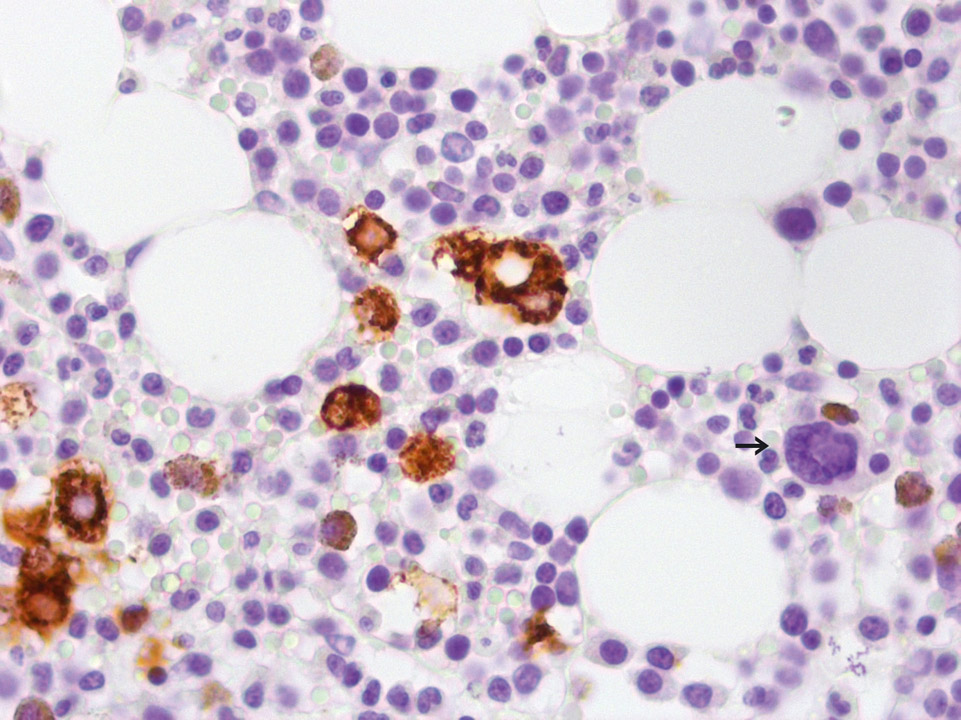
Bone marrow histology (immunohistochemistry with alkaline phosphatase reaction) of a patient with breast cancer: Carcinoma cells derive from epithelial cells that express cytokeratins in the cytoplasm. They were detected immunohistochemically with an antibody against cytokeratin 7 and visualised with a brownish stain. A megakaryocyte (->) has a size similar to the tumour cells, but is not labelled by this staining procedure.
<p>Bone marrow histology (immunohistochemistry with alkaline phosphatase reaction) of a patient with breast cancer: Carcinoma cells derive from epithelial cells that express cytokeratins in the cytoplasm. They were detected immunohistochemically with an antibody against cytokeratin 7 and visualised with a brownish stain. A megakaryocyte (->) has a size similar to the tumour cells, but is not labelled by this staining procedure.</p>
